X-Ray Imaging of Ancient Artifacts
Synchrotron-based X-ray fluorescence imaging is a powerful techique for technique for measuring and mapping elemental concentrations near the surface of an object. In collaboration with CHESS staff scientists we have been developing and applying this technique for study of a variety of ancient artifacts.
XRF Imaging of Greek and Latin Inscriptions on Stone

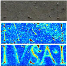
A substantial fraction of all known Greek and Latin inscriptions on stone have surfaces that are worn, weathered or abraded so that the original text is no longer visible. Previous methods for recovering this text - using shadowed light and laser scanning - were based on surface relief. We have been using synchrotron-based X-ray fluorescence imaging to search for chemical traces of inscription. Concentrations of several elements are enhanced in inscribed regions, and likely arise from wear of inscribing tools and from pigments originally used to make the letters more visible. For inscriptions from the Roman era, trace amounts of lead correlating with inscription can be seen even in regions where the surface has worn below its originally inscribed contours. These chemical traces may thus allow recovery of text for which topographic evidence has vanished, and provide information about how the inscriptions were made.
X-ray fluorescence recovers writing from ancient inscriptions. J. Powers, N. Dimitrova, R. Huang, D.-M. Smilgies, D. H. Bilderback, K. Clinton, and R. E. Thorne, Zeitschrift fur Papyrologie und Epigraphik 152, 221-227 (2005).
Dual-detector X-ray fluorescence imaging of ancient artifacts with with surface relief. D. M. Smilgies, J. A. Powers, D. H. Bilderback and R. E. Thorne. J. Synchrotron Rad. 19, 547-550 (2012).
Correcting for surface topography in X-ray fluorescence imaging. E. C. Geil and R. E. Thorne. J. Synch. Rad. 21, 1358-1363 (2014). DOI: 10.1107/S160057751401875X
X-ray fluorescence imaging and analysis of inscription provenance

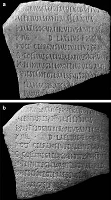
We used XRF imagining and analysis together with X-ray diffraction and electron microprobe to evaluate the provenance of a stone document from New York University strongly resembling one from Teanum Sidicium held at the American Academy in Rome. These analyses showed that the inscription was a relatively recent copy, fabricated by a different method than the original.
X-ray fluorescence imaging analysis of inscription provenance. J. Powers, D. M. Smilgies, E. C. Geil, K. Clinton, N. Dimitrova, M. Peachin and R. E. Thorne, J. Archaeological Sci. 36, 343-350 (2009).
X-ray imaging of ceramics from the American Southwest

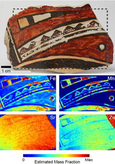
We have examined sherds of painted ceramics from prehistoric cultures of the American Southwest. Paints can generally be detected and distinguished by the fluorescence intensities of their constituent elements. Spatial maps of element distributions yield the spatial distribution of pigments. Pigments can be distinguished that are (or have become) visually similar; layers that have been obscured by overpainting can be examined; and pigment residues can be distinguished from surface contaminants deposited after painting and firing. As a result, XRFI allows the painted motifs to be clarified and hidden features to be revealed. Furthermore, the very rapid scanning and high sensitivity elemental detection possible with synchrotron-based XRFI facilitate measurements on large collections of sherds, allowing an integrative rather than anecdotal analysis.
Application of X-ray fluorescence imaging to ceramics from the American Southwest. E. C. Geil, S. A. LeBlanc, D. S. Dale and R. E. Thorne. J. Arch. Sci. 40, 4780-4784 (2013). DOI: 10.1016/j.jas.2013.05.014